New Joint Statement Streamlines Definition of Metabolic Syndrome

Sunday, October 11, 2009

by Michael O'Riordan

October 8, 2009 (Brussels, Belgium) — A new joint statement from a number of professional organizations has identified specific criteria for the clinical diagnosis of the metabolic syndrome, tightening up the definition, which previously differed from one organization to the next [1].

The statement, published online October 5, 2009 in Circulation, includes the participation of the International Diabetes Federation (IDF), the National Heart, Lung, and Blood Institute (NHLBI), the World Heart Federation, the International Atherosclerosis Society, and the American Heart Association (AHA) and is an attempt to eliminate some of the confusion regarding how to identify patients with the syndrome.

"This paper represents an attempt to make the definition global," Dr Robert Eckel (University of Colorado, Denver), one of the authors of the new report, told heartwire . "The IDF definition and the [National Cholesterol Education Program Adult Treatment Panel] ATP III definition have been the two that have been utilized most frequently, and now the different organizations--the IDF, the International Atherosclerosis Society, the NHLBI, and the AHA--have all signed on to a single definition. I think that's a step forward in terms of not continuing to confuse people who are working in this field."

Specifically, the new metabolic-syndrome definition streamlines previous differences related to abdominal obesity as defined by measurements in waist circumference. Substantial disparities existed between the previous IDF and the ATP III definitions of what constituted an excessively large waist circumference, by as much as 8 cm between the two groups, but these have been amended. Now, the criteria for elevated waist circumference are based on population- and country-specific definitions, which, although streamlined, do leave some work to be done, said Eckel.

"The problem that still exists is that regional differences around the world may be substantial in terms of what waist circumference confers additional risk for heart disease and diabetes," he said. "The new definition relies on different geographic regions, or different countries, to drill down into their own databases in terms of relating waist circumference to risk." Eckel noted that the IDF previously considered elevations in waist circumference mandatory when defining metabolic syndrome, although the ATP III did not. Now, waist circumference is just one of five criteria that physicians can use when diagnosing the metabolic syndrome. Patients with three of the five criteria--including elevated waist circumference, elevated triglycerides, reduced HDL-cholesterol levels, elevated blood pressure, and elevated fasting-glucose levels--are considered to have the syndrome.

Criteria for Clinical Diagnosis of the Metabolic Syndrome

Measure Categorical cut points
Elevated waist circumference Population- and country-specific definitions
Elevated triglycerides (drug treatment for elevated triglycerides is an alternate indicator) >150 mg/dL
Reduced HDL cholesterol (drug treatment for reduced HDL cholesterol is an alternate indicator) <40 mg/dL for males and <50 mg/dL for females
Elevated blood pressure (drug treatment for elevated blood pressure is an alternate indicator) Systolic >130 mm Hg and/or diastolic >85 mm Hg
Elevated fasting glucose (drug treatment for elevated glucose is an alternate indicator) >100 mg/dL

Notably absent from the joint statement is the American Diabetes Association. As reported by heartwire , there are unresolved scientific issues between the ADA and other associations, including the AHA, regarding the metabolic syndrome. Specifically, the ADA, as well as the European Association for the Study of Diabetes (EASD), objected to the manner in which the metabolic syndrome was characterized as a risk factor for heart disease or diabetes, arguing that there was no need to diagnose a patient with the syndrome because emphasis should be placed on aggressively treating the individual risk factors. In 2005, the ADA and EASD issued their own joint statement calling for a critical appraisal of the metabolic syndrome, its designation as a syndrome, and its clinical utility.

To heartwire , Eckel said the IDF, AHA, NHLBI, and others began working on the new metabolic syndrome definition in 2008 and that they simply went ahead without ADA participation. He stressed the metabolic syndrome is not a disease but simply a clustering of risk factors. The original intention of identifying the syndrome was simply to draw clinicians' and the public's attention to the importance of a high-quality lifestyle, and the metabolic syndrome is never meant to be used as a predictor of heart disease or diabetes risk.

References
  1. Alberti KG, Eckel RH, Grundy SM, et al. Harmonizing the metabolic syndrome. A joint interim statement of the International Diabetes Federation Task Force on Epidemiology and Prevention; National Heart, Lung, and Blood Institute; American Heart Association; World Heart Federation; International Atherosclerosis Society; and International Association for the Study of Obesity. Circulation 2009; 120:1640-1645. Abstract

Read More .......

New Joint Statement Streamlines Definition of Metabolic Syndrome

by Michael O'Riordan

October 8, 2009 (Brussels, Belgium) — A new joint statement from a number of professional organizations has identified specific criteria for the clinical diagnosis of the metabolic syndrome, tightening up the definition, which previously differed from one organization to the next [1].

The statement, published online October 5, 2009 in Circulation, includes the participation of the International Diabetes Federation (IDF), the National Heart, Lung, and Blood Institute (NHLBI), the World Heart Federation, the International Atherosclerosis Society, and the American Heart Association (AHA) and is an attempt to eliminate some of the confusion regarding how to identify patients with the syndrome.

"This paper represents an attempt to make the definition global," Dr Robert Eckel (University of Colorado, Denver), one of the authors of the new report, told heartwire . "The IDF definition and the [National Cholesterol Education Program Adult Treatment Panel] ATP III definition have been the two that have been utilized most frequently, and now the different organizations--the IDF, the International Atherosclerosis Society, the NHLBI, and the AHA--have all signed on to a single definition. I think that's a step forward in terms of not continuing to confuse people who are working in this field."

Specifically, the new metabolic-syndrome definition streamlines previous differences related to abdominal obesity as defined by measurements in waist circumference. Substantial disparities existed between the previous IDF and the ATP III definitions of what constituted an excessively large waist circumference, by as much as 8 cm between the two groups, but these have been amended. Now, the criteria for elevated waist circumference are based on population- and country-specific definitions, which, although streamlined, do leave some work to be done, said Eckel.

"The problem that still exists is that regional differences around the world may be substantial in terms of what waist circumference confers additional risk for heart disease and diabetes," he said. "The new definition relies on different geographic regions, or different countries, to drill down into their own databases in terms of relating waist circumference to risk." Eckel noted that the IDF previously considered elevations in waist circumference mandatory when defining metabolic syndrome, although the ATP III did not. Now, waist circumference is just one of five criteria that physicians can use when diagnosing the metabolic syndrome. Patients with three of the five criteria--including elevated waist circumference, elevated triglycerides, reduced HDL-cholesterol levels, elevated blood pressure, and elevated fasting-glucose levels--are considered to have the syndrome.

Criteria for Clinical Diagnosis of the Metabolic Syndrome

Measure Categorical cut points
Elevated waist circumference Population- and country-specific definitions
Elevated triglycerides (drug treatment for elevated triglycerides is an alternate indicator) >150 mg/dL
Reduced HDL cholesterol (drug treatment for reduced HDL cholesterol is an alternate indicator) <40 mg/dL for males and <50 mg/dL for females
Elevated blood pressure (drug treatment for elevated blood pressure is an alternate indicator) Systolic >130 mm Hg and/or diastolic >85 mm Hg
Elevated fasting glucose (drug treatment for elevated glucose is an alternate indicator) >100 mg/dL

Notably absent from the joint statement is the American Diabetes Association. As reported by heartwire , there are unresolved scientific issues between the ADA and other associations, including the AHA, regarding the metabolic syndrome. Specifically, the ADA, as well as the European Association for the Study of Diabetes (EASD), objected to the manner in which the metabolic syndrome was characterized as a risk factor for heart disease or diabetes, arguing that there was no need to diagnose a patient with the syndrome because emphasis should be placed on aggressively treating the individual risk factors. In 2005, the ADA and EASD issued their own joint statement calling for a critical appraisal of the metabolic syndrome, its designation as a syndrome, and its clinical utility.

To heartwire , Eckel said the IDF, AHA, NHLBI, and others began working on the new metabolic syndrome definition in 2008 and that they simply went ahead without ADA participation. He stressed the metabolic syndrome is not a disease but simply a clustering of risk factors. The original intention of identifying the syndrome was simply to draw clinicians' and the public's attention to the importance of a high-quality lifestyle, and the metabolic syndrome is never meant to be used as a predictor of heart disease or diabetes risk.

References
  1. Alberti KG, Eckel RH, Grundy SM, et al. Harmonizing the metabolic syndrome. A joint interim statement of the International Diabetes Federation Task Force on Epidemiology and Prevention; National Heart, Lung, and Blood Institute; American Heart Association; World Heart Federation; International Atherosclerosis Society; and International Association for the Study of Obesity. Circulation 2009; 120:1640-1645. Abstract

Read More .......

Pantoprazole Better Than Omeprazole for Clopidogrel-Treated Patients

NEW YORK (Reuters Health) Oct 02 - In patients with coronary stents for non-ST-segment elevation acute coronary syndrome, use of pantoprazole instead of omeprazole as a proton pump inhibitor (PPI) allows a better platelet response to clopidogrel, French researchers report.

In the September 22nd issue of the Journal of the American College of Cardiology, Dr. Thomas Cuisset of CHU Timone, Marseille, and colleagues observe that omeprazole reportedly decreases clopidogrel's antiplatelet effect, perhaps in association with CYP2C19-related mechanisms involved in PPI metabolism.

To determine whether this was a class effect, the researchers randomized 104 patients after cardiac stent placement to treatment with 20 mg of pantoprazole or 20 mg of omeprazole daily, along with dual antiplatelet therapy with 75 mg of aspirin and 150 mg of clopidogrel.

At 1 month, response to clopidogrel, as assessed by platelet reactivity index vasoactive stimulated phosphoprotein, was significantly better with pantoprazole.

Furthermore, 44% of patients in the omeprazole group were nonresponders to clopidogrel, compared to 23% of patients in the pantoprazole group.

The investigators conclude that "the degree of the interaction between clopidogrel and PPI is not homogeneous within the class of PPIs and is less marked with pantoprazole than with omeprazole."

As Dr. Cuisset told Reuters Health, "For clopidogrel-treated patients requiring a PPI," the study findings suggest "the preferential use of pantoprazole compared to omeprazole to avoid any potential negative interaction with CYP2C19."

Reference

J Am Coll Cardiol 2009;54:1149-1153.

Read More .......

Bedtime Aspirin Lowers Ambulatory Blood Pressure

By Will Boggs, MD

NEW YORK (Reuters Health) Aug 12 - Aspirin taken at bedtime reduces ambulatory blood pressure in patients with prehypertension, according to a report in the August issue of the American Journal of Hypertension.

"Bedtime ingestion of low-dose aspirin could provide a cost-effective approach for blood pressure control in subjects with prehypertension, thus delaying the actual development of hypertension and the need for costly pharmacologic treatment," Dr. Ramon C. Hermida from University of Vigo, Spain, told Reuters Health in an email.

Dr. Hermida and colleagues investigated the influence of aspirin on ambulatory blood pressure in patients who received aspirin at different times of the day according to their rest-activity cycle and who were evaluated by 48-hour ambulatory blood pressure monitoring before and after 3 months of pharmacologic intervention.

Blood pressure did not change after nonpharmacological intervention or after administration of aspirin upon awakening, the researchers note.

In contrast, the authors report, systolic blood pressure decreased by 6 mm Hg and diastolic blood pressure decreased by 3 mm Hg in patients who took 100 mg aspirin at bedtime.

Nearly three quarters (71%) of the patients who took aspirin at bedtime experienced at least a 3 mm Hg reduction in 24-hour systolic blood pressure, but only 6% showed an increase in blood pressure after treatment.

Heart rate was unchanged in all treatment groups.

"Aspirin given at the end of the activity cycle, but not upon awakening, could thus target the nocturnal peak of plasma renin activity, while enhancing the nocturnal trough in the production of nitric oxide," the investigators suggest.

"Thus," they continue, "the enhanced reduction in plasma renin activity, beneficial impact on endothelial function and blocking of alpha- and beta-adrenergic receptors associated (with) bedtime administration of low-dose aspirin could explain the impact of this intervention on blood pressure here documented in subjects with high-normal blood pressure."

"We are currently developing a new multicenter prospective trial with awakening versus bedtime administration of low-dose aspirin for primary prevention of cardiovascular events in subjects with impaired fasting glucose or diabetes," Dr. Hermida explained. "The study will involve 3200 subjects who will be followed for over 5 years."

Reference

Am J Hypertens 2009;22:896-903.

Read More .......

Left Main Artery Restenosis After Stent Placement Often Amenable to PCI

by Will Boggs, MD

NEW YORK (Reuters Health) Oct 06 - In most patients who receive drug-eluting stents for unprotected left main disease, restenosis can be managed with a minimally invasive approach, including placement of additional drug-eluting stents, according to results from the Failure in Left Main Study (FAILS).

"The FAILS study shows that repeat percutaneous coronary intervention (PCI) can be safely and effectively performed in most cases of left main restenosis," Dr. Giuseppe Biondi-Zoccai from the University of Turin, Italy told Reuters Health. "Yet, in a small but still relevant subset of patients, coronary artery bypass grafting (CABG) will still be needed, especially when restenosis is diffuse or atherosclerotic disease elsewhere in the coronary vessels is progressing rapidly."

Dr. Biondi-Zoccai and colleagues analyzed the incidence and management of restenosis in a cohort of 718 patients with unprotected left main disease who were managed with drug-eluting stents. Restenosis developed in 70 patients (9.7%).

Overall, 22.1% of cases were diagnosed during routine follow-up angiography, and 30.8% were found in patients who had been admitted for acute coronary syndrome.

According to the report in the September 22nd Journal of the American College of Cardiology, 59 of the 70 patients (84.3%) were treated again with PCI. In the repeat-PCI group, 34 patients had a new drug-eluting stent implanted, 1 received a bare metal stent, and 16 had balloon-only PCI. Six were treated with cutting balloons, and 2 underwent directional atherectomy.

Another 7 patients (10%) were managed with CABG, and 4 (5.7%) were treated with medical therapy only.

There was one in-hospital death in the repeat PCI group and none in the medical therapy or CABG groups.

Over an average of 25.6 months after restenosis, the major adverse coronary event rate was 25.7%, with death in 5.7%, myocardial infarction in 2.9%, target lesion revascularization in 21.4%, and PCI on other vessels in 21.4%. In the PCI group, there was 1 case of possible stent thrombosis, 1 case of probable stent thrombosis, and no cases of definite stent thrombosis, the investigators say.

"The bottom line of the FAILS study is that left main restenosis after PCI is not by default an indication to CABG," Dr. Biondi-Zoccai told Reuters Health. "If, in a given patient, the same reasons leading to the choice of PCI vs. CABG still hold true, than repeat PCI is feasible, safe, and can effectively manage symptoms and signs of ischemia in most subjects."

"However," Dr. Biondi-Zoccai added, "if coronary disease is extensive, restenosis is diffuse, uncontrolled diabetes mellitus is present, surgical risk is not prohibitive, and/or the patient wishes to minimize the risk of repeat revascularization, then CABG should be viewed as the treatment of choice instead of PCI for unprotected left main restenosis."

Reference

J Am Coll Cardiol 2009;54:1131-1136.

Read More .......

New ASCOT Analysis: Beta Blockers Not Beneficial in Hypertensives With Tachycardia

Sunday, October 4, 2009

by Lisa Nainggolan

September 24, 2009 — A new analysis of the Anglo-Scandinavian Cardiac Outcomes Trial--Blood Pressure Lowering Arm (ASCOT-BPLA) shows that the amlodipine (Norvasc, Pfizer)-based arm of the study remained superior to the beta-blocker arm, even when resting heart rate was taken into account [1].

The study was published in the September 22, 2009 issue of the Journal of the American College of Cardiology.

The findings mean that "there is no reason to believe that beta blockers should be used earlier on in the treatment of hypertension on the basis of heart rate," lead author Dr Neil R Poulter (Imperial College London, UK) told heartwire . He said many cardiologists have had "a hindbrain belief" that "if there is a touch of tachycardia, beta blockers are the right drugs to use in hypertension management." But prior to this analysis, there had been no data from randomized controlled trials available to assess the impact of this advice on patient outcomes, he noted.

"We thought, if it's true that a touch of tachycardia should push you toward beta blockers, we'd be able to see that in ASCOT," Poulter added. Noting that the ASCOT-BPLA study–-reported in 2005--confirmed the superiority of an amlodipine-based regimen over an atenolol-based regimen, "we looked to see whether, if you started with tachycardia, was that still the case?" he noted. "The bottom line is, yes, it is. Pulse rate should not be the determinant of what drugs you use in hypertension."

These findings "reinforce the UK guidelines that state that, in primary prevention, beta blockers should be kept to a much later stage of intervention," Poulter says.

No Attenuation of Amlodipine Superiority on Basis of Heart Rate

The ASCOT trial enrolled 19 257 hypertensive patients with at least three other cardiovascular risk factors from 650 general practices in the UK, Ireland, and the Nordic countries. The results showed that an antihypertensive strategy based on amlodipine, with perindopril added as required, significantly reduced all-cause mortality and other cardiovascular end points, including stroke, compared with an atenolol-based strategy, with the diuretic bendroflumethiazide added as required.

In this new subgroup analysis, patients with atrial fibrillation or taking rate-limiting antihypertensive drugs at baseline were excluded. The potential attenuation of the treatment effect with higher baseline heart rate on total cardiovascular events and procedures (TCVP) was assessed via an interaction term. Secondary analyses evaluated coronary and stroke outcomes.

There were 12 759 of the ASCOT patients included and 1966 total cardiovascular events and procedures. At the final visit, mean heart-rate reduction from baseline was 12.0 and 1.3 beats per minute in the atenolol- and amlodipine-based groups, respectively. There was a reduction in total cardiovascular events and procedures in those allocated to amlodipine-based therapy compared with those on atenolol-based therapy (unadjusted hazard ratio 0.81; p<0.001).

This benefit was unattenuated at higher heart rates (interaction p value=0.82). Similar results were obtained for coronary and total stroke outcomes.

"These analyses provide no evidence that atenolol-based therapy is superior to amlodipine-based therapy for patients with hypertension uncomplicated by coronary heart disease across the wide range of baseline heart rates observed in the ASCOT database," say Poulter et al.

"Pending further information--which could perhaps be gleaned from other studies such as [Losartan Intervention for Endpoint Reduction in Hypertension] LIFE--beta-blocker–based therapies are not appropriate to select as initial therapy for hypertension on the basis of a higher heart rate unless congestive heart failure and/or symptomatic ischemic heart disease coexist," they conclude.

Results Apply to All Beta Blockers, Until Further Notice

Poulter added to heartwire that "some cardiologists will say that if we hadn’t used atenolol the results would have been different: 'If you'd used my favorite "olol," ' for example, and I say, 'Prove it!' "

He believes that beta blockers "are not all the same, they do have different effects on all sorts of things," but "there is no evidence of heterogeneity in terms of cardiovascular outcomes," he maintains.

"Until further notice, beta blockers should not come up the pecking order on the basis of pulse rate."

Poulter has served as a consultant to and received travel expenses, payment for speaking at meetings, or funding for research from one or more pharmaceutical companies marketing BP-lowering or lipid-lowering drugs. Poulter has also previously received financial support from Pfizer to cover administrative and staffing costs of the ASCOT study and travel or accommodation expenses or both incurred by attending relevant meetings. Disclosures for the coauthors are listed in the paper.

References

  1. Poulter NR, Dobson JE, Sever PS, et al. Baseline heart rate, antihypertensive treatment, and prevention of cardiovascular outcomes in ASCOT (Anglo-Scandinavian Cardiac Outcomes Trial). J Am Coll Cardiol 2009; 54:1154-1161.
Read More .......

Early Statin Therapy in ACS: What's the Level of Evidence?

by Lisa Nainggolan


September 29, 2009 (Los Angeles and San Francisco, California) — A group of California cardiologists is questioning the strength of the evidence informing current US guidelines recommending the use of high-dose statin therapy early in the course of acute coronary syndrome (ACS) [1].

In a viewpoint and commentary article in the October 6, 2009 issue of the Journal of the American College of Cardiology, Dr Ryan P Morrissey (Cedars Sinai Medical Center, Los Angeles, CA) and colleagues say the current evidence is insufficient to support the class IA recommendation that is given to this indication by the ACC/AHA joint task force, a recommendation that has recently been elevated to a performance measure.

Senior author of the paper, Dr Sanjay Kaul (Cedars Sinai Medical Center), told heartwire that giving early intensive statin therapy in ACS before hospital discharge and regardless of the baseline level of LDL has been formally endorsed as a treatment guideline, "but there's never been any high-quality randomized evidence informing that recommendation." And therein lies the rub, he says, because since this advice is already given the highest recommendation, "there's no motivation for any one individual, sponsor, or professional body to pursue that kind of research, so that's a disservice."

But in a counterpoint article in the same issue [2], Drs David D Waters (San Francisco General Hospital, CA) and Ivy Ku (UCSF) argue that guidelines "prove their utility when their implementation improves outcomes across a broad population at risk, such as in this instance." Commenting on the paper by Morrissey et al, they say: "We disagree with their conclusions and with their interpretation of the facts."

Waters told heartwire : "Statins are the biggest pharmacologic advance we've ever had in cardiology to treat coronary disease. After an acute coronary event, the risk of another event, another MI, hospitalization for unstable angina, or cardiac death is highest in the first six months, and these drugs have been shown in two trials--MIRACL and PROVE-IT--to reduce events at that time." Waters says Morrissey et al are "nitpicking. It's hard to say much about their paper without sounding uncharitable."

Strengths and Limitations to the Evidence

The basic thrust of the argument by the Cedars Sinai doctors is that the current evidence from randomized trials--while indicating that early initiation of high-dose statin therapy in ACS does reduce recurrent ischemia and may reduce revascularization--does not indicate benefit in terms of hard clinical outcomes such as death or MI and may be associated with increased liver and muscle-related adverse outcomes.

"Our message should not be that patients should not be treated with statins," Kaul stressed to heartwire . "Our message is that statins are useful and effective drugs, but you have to recognize that there are strengths and limitations to the evidence."

Kaul says that "from a practical perspective, the evidence is sufficient for clinical practice," but "there are many things we do in clinical practice that are not well-informed by high-quality evidence. . . . That does not necessarily invalidate them. But strictly speaking, from a technical perspective, the evidence does not support a class IA recommendation for this indication. Even though we tend to conflate the two, 'general agreement' should not have the same currency as 'evidence.' Guidelines should dictate the 'standard of care,' not be driven by them."

As substance for his argument, he points out that the European guidelines on this "are somewhat more faithful to the evidence. They say to start early, within one to four days of ACS admission, but this is given a class IB rather than a class IA recommendation, and they put their target treatment level at <100 mg/dL. For the more aggressive treatment target of 70 mg/dL, they have a class IIA, level B recommendation," he notes, adding, "This is more in line with the evidence than the American guidelines. So here we have two professional societies looking at the same evidence and coming up with different recommendations--it's open to interpretation and it becomes more of an opinion."

Kaul also objects to the fact that the recommendations to begin high-dose statin therapy early in the course of ACS have been elevated to a performance measure "using the same evidence. Now our performance is going to be judged on this, and that has broad implications."

Statins Early in ACS a "No-Brainer"

For his part, Waters says, "it's very hard to get a clinical trial to show benefit now because there's so much standard treatment that's beneficial. These two trials [MIRACL and PROVE-IT] clearly show you can reduce end points like stroke, heart attack, hospitalization for recurrent angina, and coronary revascularization with statins.

"It's kind of a no-brainer. The guidelines pretty much universally have recommended that patients with ACS take these drugs, and I think it's made a big difference."

As to the discordance between the US and European guidelines, Waters says: "It's true, there is a slight discordance. But anywhere you get two august bodies of experts on different sides of the ocean trying to turn the results of trials into recommendations for treatment, it's a little tricky."

He adds: "We have more trials with statins in cardiology than we do with any other drug class. We've got randomized, double-blind, placebo-controlled trials in well over 100 000 patients, and we haven't yet found a level below which lowering your LDL threshold doesn't provide additional benefit. If you lower LDL, you get benefits; some say it's from effects other than LDL lowering, and that may be true."

Kaul Would Take a Statin for ACS, But Personal Choice and Policy Don't Mix

Asked by heartwire whether he would personally take a statin early in the course of ACS were he unfortunate enough to suffer such an event, Kaul said: "Yes, I would definitely start with a moderate-dose statin and be content with an on-treatment LDL level of 75 or 80 mg/dL." However, he notes there is "nothing magical about an LDL threshold of 70" and says that starting on a modest dose would "enable me to avoid the side effects, thereby minimizing the potential for nonadherence and maximizing the likelihood of benefit."

But Kaul stresses that his above response in no way invalidates what he and his colleagues are trying to say in their paper. "We need to separate personal belief or clinical judgment from policy statements that have broad implications. Guidelines work best when they are based on empirical facts, not cogent hypotheses; when they inform our clinical judgment, not replace it. Guidelines that are not informed by high-quality, strong evidence have the potential to misinform future research and lead to suboptimal clinical practice.

"I want to emphasize that the goal of our paper is to make people more aware of the quality of the evidence that informs the guidelines, and hopefully the [National Cholesterol Education Program Adult Treatment Panel] NCEP ATP IV recommendations coming out in the near future will be more cognizant of the strengths and weaknesses of the data and will implement their recommendations accordingly," he concludes.

Waters has received honoraria for lectures from Pfizer and consulting fees from AstraZeneca, Merck/Schering-Plough and Pfizer. Kaul reports no conflicts of interest; coauthor Dr George A Diamond (Cedars Sinai Medical Center) serves on the speaker's panel of Schering-Plough and Merck.

References

  1. Morrissey RP, Diamond GA, Kaul S et al. Statins in acute coronary syndromes. Do the guidelines match the evidence? J Am Coll Cardiol 2009; 54:1425-1433.
  2. Waters DD and Ku I. Early statin therapy in acute coronary syndromes. The successful cycle of evidence, guidelines and implementation. J Am Coll Cardiol 2009; 54:1434-1437.
Read More .......

Doubling Dose of Clopidogrel Benefits STEMI Patients Without Risk of Bleeding

by Michael O’Riordan

September 25, 2009 (San Francisco, California) — Doubling the loading and maintenance dose of clopidogrel (Plavix, Bristol-Myers Squibb/Sanofi-Aventis) in ST-segment-elevation-MI (STEMI) patients reduces ischemic events and stent thrombosis, without significantly increasing the risk of bleeding, a new analysis shows. The reduction in stent thrombosis occurred in patients treated with bare-metal stents and drug-eluting stents, according to investigators.

These are the results of a PCI substudy from the Clopidogrel Optimal Loading Dose Usage to Reduce Recurrent Events/Optimal Antiplatelet Strategy for Interventions (CURRENT OASIS-7) study. The postrandomization analysis, dubbed CURRENT STEMI PCI, looked specifically at patients undergoing PCI for STEMI and is in line with results of all patients with acute coronary syndrome (ACS) who underwent PCI. In contrast to the ACS findings, there was no increased need for red blood cell transfusions with STEMI patients.

"The reason for this, I suppose, is that one of the biggest drivers of bleeding in an ACS cohort is age, and the STEMI patients tended to be younger," said lead investigator Dr Shamir Mehta (McMaster University, Hamilton, ON) during a morning press conference here at the TCT 2009 meeting. "The mean age of the STEMI patients was 59 years, whereas those without STEMI tended to be older, have more comorbidities, and tended to have higher rates of bleeding. This has been seen in other trials."

Commenting on the results of the study during the late-breaking clinical-trials session, moderator Dr Gregg Stone (Columbia University, New York) called the CURRENT STEMI PCI an important trial that should have an immediate impact on clinical practice. While interventionalists at Columbia were already using a 600-mg loading dose of clopidogrel, he said his group will likely begin using a 150-mg maintenance dose in the seven days following the procedure, as was the protocol in the study. Panel member Dr Cindy Grines (Beaumont Hospital, Royal Oak, MI) agreed, telling the audience that her group would follow the clopidogrel loading and maintenance protocol, while Dr Gilles Montalescot (Institut de Cardiologie, Pitié-Salpêtrière Hospital, Paris, France) said his hospital has already doubled the dose in the ACS setting.

CURRENT STEMI PCI Cohort of CURRENT OASIS-7

The overall results of the CURRENT OASIS-7 study were previously reported by heartwire , but briefly, the study is a 2x2 factorial, randomized trial studying the optimal doses of clopidogrel and aspirin in ACS patients presenting to an emergency department and scheduled to undergo an early invasive strategy with intent to perform PCI no later than 72 hours after randomization. Of the original 17 232 patients undergoing PCI, 6346 patients presented with STEMI, a significant majority of whom underwent primary PCI.

Patients assigned to high-dose clopidogrel received a 600-mg loading dose on day 1 and then 150 mg once daily for next seven days, followed by 75 mg once daily until 30 days. Patients in the standard clopidogrel arm received a 300-mg loading dose on day 1, followed by 75 mg once daily until 30 days. Patients were also assigned in an open-label manner to 300 to 325 mg of aspirin once daily or 75 to 100 mg aspirin once daily.

At 30 days, there was a significant reduction in the risk of ischemic events, specifically a 37% reduction in MI, in patients who received the double dose of clopidogrel. In addition, there was a significant 46% reduction in the risk of definite stent thrombosis and a 30% reduction in risk in the combined end point of MI and all stent thrombosis. There was no increased risk of bleeding.

CURRENT STEMI PCI: Efficacy Outcomes

Outcome Standard clopidogrel (n=3175) Double-dose clopidogrel (n=3171) Hazard ratio (95% CI)
Definite stent thrombosis 1.8 1.0 0.54 (0.35–0.84)
All stent thrombosis 3.5 2.5 0.72 (0.54–0.96)
MI 1.9 1.2 0.63 (0.41–0.94)
MI or stent thrombosis 4.0 2.8 0.70 (0.54–0.92)
Cardiovascular death 3.2 3.1 0.95 (0.72–1.26)
Stroke 0.5 0.4 0.81 (0.39–1.69)

CURRENT STEMI PCI: Bleeding

Bleeding definition Standard clopidogrel (n=3175) Double-dose clopidogrel (n=3171) Hazard ratio (95% CI)
CURRENT major 1.2 1.4 1.16 (0.75–1.78)
CURRENT severe 0.9 1.1 1.18 (0.72–1.93)
Fatal 0.3 0.1 0.33 (0.09–1.23)
ICH 0.03 0.03 1.00 (0.06–16.01)
Red blood cell transfusion >2U 1.0 1.1 1.20 (0.74–1.95)
CABG-related major 0.03 0.2 6.01 (0.72–49.88)

ICH=intracranial hemorrhage

Discussing the results with the media and during the late-breaking clinical-trials session, Mehta said that the benefit of double-dose clopidogrel was observed early and that there was no increased risk, mainly in stent thrombosis catch-up, when clinicians switched from 150-mg to 75-mg of clopidogrel on day 7. The reduction in risk of definite stent thrombosis was significant in patients who received a bare-metal stent (37% reduction in risk) or a drug-eluting stent (50% reduction in risk).

Despite the praise of the study, some researchers were cautious in their interpretation, noting that the overall results were "flat" and that there was a significant interaction with aspirin. As reported previously by heartwire , CURRENT OASIS-7 included 7855 patients who did not undergo PCI because no significant CAD was identified on the coronary angiogram or who discontinued therapy because they were scheduled for CABG surgery, and when these were included in the overall analysis, there was no significant difference in the primary composite end point of death, MI, or stroke between the two treatment arms.

Dr Robert Harrington (Duke Clinical Research Institute, Durham, NC) noted that this STEMI analysis was a postrandomization cohort and included all patients who underwent primary PCI, rather than those who were simply randomized to undergo the intervention. Dr Ajay Kirtane (Columbia University, New York) agreed.

"This is a postrandomization subgroup in an overall negative trial," said Kirtane. "I think we should be cautious in overinterpreting this. I think it's a very difficult question. Pathophysiologically, increased antiplatelet inhibition should theoretically lower thrombotic events, but it's a difficult conundrum we're in with this trial."

Responding to the questions, Mehta said that the intention-to-treat analysis also showed significant reductions in stent thrombosis and MI. He also said that CURRENT OASIS-7 was not "negative" because of the significant interaction with aspirin. In a 2X2 factorial designed study, if there is an interaction, researchers should look at the study arms separately, he said, and when they did this, there was a significant overall reduction in the primary end point with the double dose of clopidogrel.

Read More .......

When and How to Implement Basal-Bolus Therapy: Treating to Success

Tuesday, September 22, 2009

by Russell D. White, MD

Type 2 Diabetes Is a Progressive Disease

Because of the progressive nature of type 2 diabetes, the majority of patients with longstanding disease are likely to be candidates for insulin therapy.[1]

  • This is due to the fact that a large fraction of β-cell function has been lost by the time the disease is diagnosed and this decline continues over time.[2]
  • During the early stages of type 2 diabetes, lifestyle interventions may be successful for improving glycemic control for some patients,[3] but as β-cell function continues to decline, pharmacologic treatment is likely to be necessary:
    • Oral therapy may progress from treatment with a single agent to combination therapy with 2 or 3 drugs.[4]
    • When oral agents are not effective in reaching A1C targets, addition of insulin therapy is required.[5]
    • There is an emerging awareness that using insulin earlier in the course of the disease is physiologically sound and should be an integral part of adequate diabetes management.[6]

    Progressive Disease Requires Progressive Treatment

    Insulin treatment for patients with diabetes should be aimed at treating to target, using information gained from careful blood glucose (BG) monitoring.[7]

    • As the disease progresses, so should therapy.
    • Treatment should be aimed at a specific A1C goal and adjusted as needed to reach it.
    • This approach is in contrast to a slower stepwise approach likely to result in repeated failure and loss of glycemic control.

    Basal Insulin

    There are multiple approaches to the initiation of insulin therapy in a patient with type 2 diabetes who can no longer maintain glycemic control on oral agents.

    • Addition of basal insulin therapy to oral drugs:
      • This approach is consistent with results from clinical trials that have shown that adding basal insulin to ongoing oral therapy can improve glycemic control.[8]
      • Addition of basal insulin is thought to improve control over BG by suppression of overnight hepatic glucose production, both through direct effects on the liver and indirect effects due to inhibition of free fatty acid release by adipose tissue.[9] Decreasing elevated fasting plasma glucose may decrease the risk for long-term complications of diabetes.[10]
      • Main options for basal insulin therapy[11]:
        • Intermediate-acting neutral protamine Hagedorn (NPH) insulin
        • Long-acting insulin analog (insulin glargine or insulin detemir)
        • The pharmacokinetic/pharmacodynamic profiles for NPH insulin and long-acting insulin analogs are substantially different. Those for the long-acting insulin analogs are relatively flat, whereas those for NPH insulin have a distinct peak at approximately 4 hours. This difference results in different dosing requirements for these insulins[9]:
          • NPH insulin must be given in the evening or at bedtime and often requires twice daily administration, with a second dose in the morning.
          • Long-acting insulin analogs can be administered at bedtime, before dinner, or in the morning, due to their longer durations of action and lower peak effects.
        • There are other important differences between NPH insulin and long-acting insulin analogs:
          • The pharmacokinetic effects of NPH insulin are more variable than those of insulin glargine or insulin detemir. This variability is a result of the manner in which NPH insulin is structured[12]:
            • Protamine is added to regular insulin to extend its duration of action. Addition of protamine increases the tendency of insulin molecules to remain in a hexameric structure at the injection site, resulting in a longer duration of action and longer time to peak. However, the resulting poor solubility of this insulin preparation increases the variability in its pharmacokinetic profile.
            • The requirement for resuspension of NPH before injection may also contribute to inter- and intrapatient variability in the action of this insulin because the actual amount of insulin administered may vary from one injection to the next.[12]
            • Long-acting insulin analogs have much less variability than NPH. The coefficient of variation for a glucose infusion in patients given 0.4 U/kg NPH insulin is 68%, that for insulin glargine is 48%, and that for insulin detemir is 27%.[13]
            • This may contribute to the lower risk for hypoglycemia with these agents versus NPH insulin.[7]
            • Clinical studies have demonstrated the effectiveness of adding a long-acting basal insulin analog to oral therapy in patients with type 2 diabetes.[7,14–16]
            • Insulin detemir appears to be associated with less weight gain than either insulin glargine or NPH insulin.[17,18]
      • How to dose basal insulin:
        • The initial dose for insulin detemir is usually 10 U or 0.1-0.2 U/kg.[19]
        • In the PREDICTIVE 303 study patients self-adjusted their insulin detemir dose every 3 days based on the average of three self-monitored BG (SMBG) values. Insulin detemir doses were adjusted as follows[20]:
          • If the mean fasting plasma glucose (FPG) is <80 mg/dL, the dose is reduced by 3 U.
          • If the mean FPG is 80 to 110 mg/dL, there is no change in dosing.
          • If the mean FPG is >110 mg/dL, the dose is increased by 3 U.
        • One recent study has demonstrated that patients can safely self-titrate insulin detemir to an FPG target of 70-90 mg/dL with low rates of hypoglycemia.[21]
        • The typical initial dose for insulin glargine is 10 U.[22] In one study, doses for insulin glargine were adjusted as follows[23]:
          • No change is required if FPG remains between 70 and 94 mg/dL.
          • If FPG is <70 mg/dL for 3 days, the dose should be decreased by up to 10% of the total dose.
          • If FPG is 95 to 119, 120 to 139, 140 to 180, or >180 mg/dL for 3 days, the dose should be increased by 2, 4, 6, or 8 U, respectively.

    Adding Prandial Insulin

    While addition of basal therapy is highly effective in many patients, the progressive nature of diabetes may require further intensification of treatment. Rapid-acting insulin analogs are a suitable first choice for intensification of therapy:

    • Rapid-acting insulin analogs have lower variability in absorption and more consistent pharmacodynamic profiles than regular human insulin.[24,25]
    • Rapid-acting insulin analogs provide higher 1- and 2-hour insulin values, reduced risk for late postprandial hypoglycemia due to a shorter duration of action than regular human insulin, and may provide quality of life benefits due to greater flexibility in timing and meal-time dosing.[26]
    • These insulin analogs provide a more physiologic action which coincides with meal patterns.

    Adding prandial insulin to basal insulin may be the best way to restore postprandial and overall glycemic control when the combination of basal insulin and oral therapies is no longer effective. Prandial insulin may be added to basal insulin in a step-wise manner[9]:

    • Adding prandial insulin to decrease postprandial glucose (PPG) may be most beneficial in patients with FPG that is at or near goal, but who still have modestly elevated A1C. Results from Monnier and colleagues have shown that PPG makes the greatest contribution to A1C at values below approximately 8.5%.[27] Addition of prandial insulin should also be considered when the basal insulin dose is very high (>0.6 U/kg).
    • A single mealtime prandial injection of a rapid-acting insulin analog that controls the highest postprandial glucose (PPG) may be sufficient to restore glycemic control[28]:
      • The basal insulin dose should be lowered by the amount of prandial insulin to decrease the risk for nocturnal hypoglycemia.[29]
      • The prandial insulin dose can then be titrated in accordance with SMBG measured 2 hours after the start of the meal, before the next meal, or at bedtime if the injection is administered before the evening meal:
        • The American Diabetes Association goal for peak PPG is <180 mg/dL.[30]
        • The American College of Endocrinology recommends a 2-hour postprandial BG level <140 mg/dL.[31]
      • It has also been shown that prandial insulin doses can be effectively adjusted based on premeal BG patterns from the previous week.[23]
      • The prandial insulin dose can be adjusted independently to limit postprandial hyperglycemia without affecting basal insulin action.[9,27]

    Basal-Bolus Therapy

    Basal-bolus therapy may be an appropriate treatment progression for patients who have had prandial insulin added to the treatment regimen[32]:

    • In these regimens, basal and bolus insulin requirements are each approximately 50% of the total daily insulin needed.
    • Results from the PREFER study indicated that one-third of the total prandial insulin dose can be delivered with each meal with the majority of patients achieving A1C ≤7%.[33]
    • It has also been suggested that the total dose of rapid-acting insulin analog may be divided with 38% delivered at breakfast, 28% at lunch, and 33% at dinner (White RD, et al., unpublished data). The highest prandial dose is delivered at breakfast for two reasons:  
      • High carbohydrate content for this meal
      • The “dawn phenomenon,” a morning surge in plasma glucose that occurs secondary to a physiologic morning rise in cortisol and growth hormone levels.[34]

    There are several considerations for titration of prandial insulin:

    • Rapid-acting insulin analogs have accelerated pharmacokinetics compared with long-acting insulins, and they require different adjustments that can be made at shorter intervals.
    • SMBG should be carried out at least three times per day for patients using multiple insulin injections.[30]
    • Exercise improves insulin sensitivity, and it is often necessary to reduce the prandial insulin before exercise occurring within 3 hours in patients who engage in moderate or strenuous exercise.[35] Carbohydrate counting is an alternative to dosage algorithms for determining prandial insulin dose, and it provides for more flexibility in meal planning,[23] but this is not absolutely necessary.
    • Concern about hypoglycemia is a significant psychological barrier to intensive insulin therapy[36]:  
      • This risk is reduced by the use of long- and rapid-acting insulin analogs versus human insulins.
      • In one study, switching patients from the combination of regular human insulin and NPH insulin to insulin lispro and insulin glargine resulted in a 44% reduction in the occurrence of hypoglycemia.[37]
      • A second study indicated a 43% reduction in the occurrence of nighttime hypoglycemia with the insulins lispro and glargine versus regular human and NPH insulin.[38]
      • The combination of insulins detemir and aspart has also been shown to be associated with 38% lower risk for nighttime hypoglycemia than NPH plus regular human insulin in patients receiving basal-bolus treatment.[39]

    Simplifying Basal-Bolus Therapy

    Advances in therapy may help to simplify basal-bolus therapy:

    • Rapid-acting insulin analogues that can be administered shortly before or even after meals[40] have the potential to improve adherence.
    • Adherence to treatment in difficult-to-manage patients may also be improved with intensive home- and community-based family therapy.[41]
    • Patient referral to specialists for intensive diabetes care may be necessary to reach treatment targets in some patients.[42]
    • Use of an insulin pen may improve adherence to treatment and thus glycemic control. It has been shown that patients with type 2 diabetes prefer insulin pen devices over syringe and vial; and these devices improve adherence to therapy and decrease the risk for hypoglycemia.[43]
    • Additional steps that may increase the probability of reaching treatment goals include[44]:
      • Monitoring A1C every 3 months in addition to SMBG
      • Aggressively managing hyperglycemia, dyslipidemia, and hypertension with the same intensity to obtain the best patient outcome
      • Implementing a multi- and interdisciplinary team approach to diabetes management both to encourage patient education and self-care and to share responsibility for patients achieving glucose goals

    References

    1. Wright A, Burden AC, Paisey RB, Cull CA, Holman RR, for the U.K. Prospective Diabetes Study Group. Sulfonylurea inadequacy. Efficacy of addition of insulin over 6 years in patients with type 2 diabetes in the U.K. Prospective Diabetes Study (UKPDS 57). Diabetes Care. 2002;25:330-336.
    2. DeFronzo RA. From the triumvirate to the ominous octet – a new paradigm for the treatment of T2DM. The Banting Lecture. Programs and abstracts of the American Diabetes Association 68th Scientific Sessions; June 6-10, 2008; San Francisco, California.
    3. Petersen KF, Dufour S, Befroy D, Lehrke M, Hendler RE, Shulman GI. Reversal of nonalcoholic hepatic steatosis, hepatic insulin resistance, and hyperglycemia by moderate weight reduction in patients with type 2 diabetes. Diabetes. 2005;54:603-608.
    4. Bell DS. Type 2 diabetes mellitus: what is the optimal treatment regimen? Am J Med. 2004;116(suppl 5A):23S-29S.
    5. Nathan DM, Buse JB, Davidson MB, et al. Medical management of hyperglycemia in type 2 diabetes: a consensus algorithm for the initiation and adjustment of therapy. A consensus statement of the American Diabetes Association and the European Association for the Study of Diabetes. Diabetes Care. 2009;32:193-203.
    6. Eldor R, Stern E, Milicevic Z, Raz I. Early use of insulin in type 2 diabetes. Diabetes Res Clin Pract. 2005;68(suppl 1):S30-S35.
    7. Riddle MC, Rosenstock J, Gerich J; on behalf of the Insulin Glargine 4002 Study Investigators. The treat-to-target trial. Randomized addition of glargine or human NPH insulin to oral therapy of type 2 diabetic patients. Diabetes Care. 2003;26:3080-3086.
    8. Davies M, Evans R, Storms F, Gomis R, Khunti K; on behalf of the AT.LANTUS Study Group. Initiation of insulin glargine in suboptimally controlled patients with type 2 diabetes: sub-analysis of the AT.LANTUS trial comparing treatment outcomes in subjects from primary and secondary care in the UK. Diabetes Obes Metab. 2007;9:706-713.
    9. Raccah D, Bretzel RG, Owens D, Riddle M. When basal insulin therapy in type 2 diabetes mellitus is not enough–what next? Diabetes Metab Res Rev. 2007;23:257-264.
    10. Gimeno-Orna JA, Castro-Alonso FJ, Boned-Juliani B, Lou-Arnal LM. Fasting plasma glucose variability as a risk factor of retinopathy in Type 2 diabetic patients. J Diabetes Complications. 2003;17:78-81.
    11. Scholtz HE, Pretorius SG, Wessels DH, Becker RH. Pharmacokinetic and glucodynamic variability: assessment of insulin glargine, NPH insulin and insulin ultralente in healthy volunteers using a euglycaemic clamp technique. Diabetologia. 2005;48:1988-1995.
    12. Takiya L, Dougherty T. Pharmacist’s guide to insulin preparations: a comprehensive review. Pharmacy Times CE. 2005. Available at: https://secure.pharmacytimes.com/lessons/200510-03.asp.
    13. Heise T, Nosek L, Rønn BB, et al. Lower within-subject variability of insulin detemir in comparison to NPH insulin and insulin glargine in people with type 1 diabetes. Diabetes. 2004;53:1614-1620.
    14. Garber AJ, Clauson P, Pedersen CB, Kølendorf K. Lower risk of hypoglycemia with insulin detemir than with neutral protamine Hagedorn insulin in older persons with type 2 diabetes: a pooled analysis of phase III trials. J Am Geriatr Soc. 2007;55:1735-1740.
    15. Fritsche A, Schweitzer MA, Häring HU; and the 4001 Study Group. Glimepiride combined with morning insulin glargine, bedtime neutral protamine Hagedorn insulin, or bedtime insulin glargine in patients with type 2 diabetes. A randomized, controlled trial. Ann Intern Med. 2003;138:952-959.
    16. Hermansen K, Davies M, Derezinski T, Martinez Ravn G, Clauson P, Home P; on behalf of the Levemir Treat-to-Target Study Group. A 26-week, randomized, parallel, treat-to-target trial comparing insulin detemir with NPH insulin as add-on therapy to oral glucose-lowering drugs in insulin-naive people with type 2 diabetes. Diabetes Care. 2006;29:1269-1274.
    17. Rosenstock J, Davies M, Home PD, Larsen J, Koenen C, Schernthaner G. A randomised, 52-week, treat-to-target trial comparing insulin detemir with insulin glargine when administered as add-on to glucose-lowering drugs in insulin-naive people with type 2 diabetes. Diabetologia. 2008;51:408-416.
    18. Fakhoury W, Lockhart I, Kotchie RW, Aagren M, LeReun C. Indirect comparison of once daily insulin detemir and glargine in reducing weight gain and hypoglycaemic episodes when administered in addition to conventional oral anti-diabetic therapy in patients with type-2 diabetes. Pharmacology. 2008;82:156-163.
    19. Levemir. [prescribing information]. 2007. Available at: http://www.levemir-us.com/prescribing_information.pdf. Accessed May 21, 2009.
    20. Selam JL, Koenen C, Weng W, Meneghini L. Improving glycemic control with insulin detemir using the 303 algorithm in insulin naïve patients with type 2 diabetes: a subgroup analysis of the US PREDICTIVE 303 study. Curr Med Res Opin. 2008;24:11-20.
    21. Blonde L, Merilainen M, Karwe V, Raskin P; TITRATE Study Group. Patient-directed titration for achieving glycaemic goals using a once-daily basal insulin analogue: an assessment of two different fasting plasma glucose targets - the TITRATE study. Diabetes Obes Metab. 2009;11:623-631.
    22. Lantus. [prescribing information]. 2007. Available at: http://products.sanofi-aventis.us/lantus/lantus.html. Accessed May 21, 2009.
    23. Bergenstal RM, Johnson M, Powers MA, et al. Adjust to target in type 2 diabetes: comparison of a simple algorithm with carbohydrate counting for adjustment of mealtime insulin glulisine. Diabetes Care. 2008;31:1305-1310.
    24. Becker RH, Frick AD. Clinical pharmacokinetics and pharmacodynamics of insulin glulisine. Clin Pharmacokinet. 2008;47:7-20.
    25. Guerci B, Sauvanet JP. Subcutaneous insulin: pharmacokinetic variability and glycemic variability. Diabetes Metab. 2005;31:4S7-4S24.
    26. Rossetti P, Porcellati F, Bolli GB, Fanelli CG. Prevention of hypoglycemia while achieving good glycemic control in type 1 diabetes. The role of insulin analogs. Diabetes Care. 2008;31(suppl 2):S113-S120.
    27. Monnier L, Lapinski H, Colette C. Contributions of fasting and postprandial plasma glucose increments to the overall diurnal hyperglycemia of type 2 diabetic patients Variations with increasing levels of HbA(1c). Diabetes Care. 2003;26:881-885.
    28. Monnier L, Colette C. Addition of rapid-acting insulin to basal insulin therapy in type 2 diabetes: indications and modalities. Diabetes Metab. 2006;32:7-13.
    29. Meneghini L. Why and how to use insulin therapy earlier in the management of type 2 diabetes. South Med J. 2007;100:164-174.
    30. American Diabetes Association. Standards of medical care in diabetes–2009.Diabetes Care. 2009;32(suppl 1):S13-S61.
    31. American College of Endocrinology. American College of Endocrinology Consensus Statement on Guidelines for Glycemic Control. Endocrinology. 2002;8(suppl 1):6-11.
    32. Mayfield JA, White RD. Insulin therapy for type 2 diabetes: rescue, augmentation, and replacement of beta-cell function. Am Fam Physician. 2004;70:489-500.
    33. Liebl A, Prager R, Binz K, Kaiser M, Bergenstal R, Gallwitz B; PREFER Study Group. Comparison of insulin analogue regimens in people with type 2 diabetes mellitus in the PREFER Study: a randomized controlled trial. Diabetes Obes Metab. 2009;11:45-52.
    34. Carroll MF, Schade DS. The dawn phenomenon revisited: implications for diabetes therapy. Endocr Pract. 2005;11:55-64.
    35. Banting and Best Diabetes Centre. Approach to the Management of Diabetes Mellitus. 6th ed. Developed by the Diabetes Care and Education Committee, Banting and Best Diabetes Centre. Toronto: Faculty of Medicine, University of Toronto; 2005.
    36. Hartman I. Insulin analogs: impact on treatment success, satisfaction, quality of life, and adherence. Clin Med Res. 2008;6:54-67.
    37. Ashwell SG, Amiel SA, Bilous RW, et al. Improved glycaemic control with insulin glargine plus insulin lispro: a multicentre, randomized, cross-over trial in people with Type 1 diabetes. Diabet Med. 2006;23:285-292.
    38. Murphy NP, Keane SM, Ong KK, et al. Randomized cross-over trial of insulin glargine plus lispro or NPH insulin plus regular human insulin in adolescents with type 1 diabetes on intensive insulin regimens. Diabetes Care. 2003;26:799-804.
    39. Raslová K, Bogoev M, Raz I, Leth G, Gall MA, Hâncu N. Insulin detemir and insulin aspart: a promising basal-bolus regimen for type 2 diabetes. Diabetes Res Clin Pract. 2004;66:193-201.
    40. Garg SK, Rosenstock J, Ways K. Optimized basal-bolus insulin regimens in type 1 diabetes: insulin glulisine versus regular human insulin in combination with basal insulin glargine. Endocr Pract. 2005;11:11-17.
    41. Ellis DA, Frey MA, Naar-King S, Templin T, Cunningham P, Cakan N. Use of multisystemic therapy to improve regimen adherence among adolescents with type 1 diabetes in chronic poor metabolic control. A randomized controlled trial. Diabetes Care. 2005;28:1604-1610.
    42. Graber AL, Elasy TA, Quinn D, Wolff K, Brown A. Improving glycemic control in adults with diabetes mellitus: shared responsibility in primary care practices. South Med J. 2002;95:684-690.
    43. Goldstein HH. Pen devices to improve patient adherence with insulin therapy in type 2 diabetes. Postgrad Med. 2008;120:172-179.
    44. Del Prato S, Felton AM, Munro N, et al; on behalf of the Global Partnership for Effective Diabetes Management. Improving glucose management: ten steps to get more patients with type 2 diabetes to glycaemic goal. Int J Clin Pract. 2005;59:1345-1355.
Read More .......

Rethinking Hypertension in the 21st Century: An Overview of the Expanded Definition and Classification of Hypertension

by Thomas D. Giles, MD

Introduction

As epidemiologic and clinical data regarding the relationship between blood pressure (BP) and the risk for cardiovascular disease (CVD) have accumulated, a pronounced shift has taken place in how the disease of hypertension is viewed and defined. Cardiovascular (CV) risk has been found to be elevated at BP levels previously considered normal; in some cases, sporadic elevations in BP levels may be physiologically benign and not associated with additional CVD risk.[1-3] As a consequence, many hypertension experts consider elevated BP at its core a disease marker, rather than a cause of hypertension. Moreover, elevated BP, as 1 marker of CVD, frequently coexists with other equally compelling disease markers.[2] Elevated BP should not, therefore, be viewed or treated in isolation, but considered in the context of whole patient care, which takes into account the presence of other risk factors and disease markers for CVD to achieve a more comprehensive, or global, assessment of CV risk.

With these points in mind, in 2005, the Hypertension Writing Group (HWG), a national group of hypertension specialists, proposed a new definition of hypertension as "a progressive cardiovascular syndrome, the early markers of which may be present even before BP elevations are observed."[4] The stated goal of the new definition was to identify individuals at risk for CVD at an earlier point in the disease process, as well as to avoid labeling persons as hypertensive who are at low risk for CVD.[4] Viewed from this perspective, the HWG believed that threshold-based classification systems of hypertension, such as that endorsed in the Seventh Report of the Joint National Committee on Prevention, Detection, Evaluation, and Treatment of High Blood Pressure (JNC 7),[5] while serving as tools to identify patients across a broad range of CVD risk, may lead to underestimation or overestimation of clinical risk within individual patients. In either case, the presence or absence of other disease markers or risk factors, the coexistence of target-organ damage, or both can be used to risk-stratify patients with hypertension more accurately.

To simplify risk stratification and align it more closely with clinical practice, the HWG proposed classifying all patients as either normal or hypertensive (eliminating the prehypertension category proposed in JNC 7), with hypertension classified into stage 1, stage 2, or stage 3.[4] Because the CV syndrome represented by hypertension may be present even when BP falls within the normal category by conventional standards, the risk categories created by the HWG focus not on BP levels per se, but on the presence of deleterious BP patterns or the presence of CVD. Stages of hypertension are further categorized based on the presence of risk factors for early, advanced, or progressive CVD, as well as by other CVD markers (classified as BP, cardiac, vascular, renal, and retinal changes) and target-organ damage (classified as cardiac, vascular, renal, and cerebrovascular).[4]

Beyond the goal of providing a more clinically relevant assessment of global CV risk in clinical practice, this paradigm shift served to focus attention on the enormous unmet need regarding prevention and optimal treatment of hypertension across a spectrum of fields, from basic research and drug development to patient education and clinical management.[4] Two critical areas of research in particular -- the development of specific and sensitive cost-effective tests that can detect early CVD markers in the clinical setting, and the development of strategies to slow or prevent the onset of target-organ damage or overt CVD by treating early vascular derangements -- may benefit from being examined within the context of the categories for hypertension.

Recently, the HWG further refined and updated the definition and classification of hypertension.[6] This article reviews the revised definition and classification scheme and the implications for clinical practice. As the authors stressed, however, while definitions of disease are useful for detection, management, research, and education, definitions alone do not constitute recommendations for treatment. In the latter case, the initiation of treatment should be individualized and guided by CV risk, rather than BP thresholds.[1]

Blood Pressure as a Biomarker for Hypertension

The concept of elevated BP as a disease marker for hypertension, rather than its cause, is supported by multiple lines of evidence suggesting that the risk for renovascular and CV sequelae may be higher than expected in the presence of normal or near-normal BP in some patients, or, conversely, lower than expected in the presence of above-normal BP in others. This view is based, in part, on the physiologically dynamic nature of BP, in which tissue perfusion is matched with metabolic demands in a complex, ever-changing manner that depends on the coordinated activity of numerous mechanisms involved in hemostasis, including the sympathetic nervous system, the renin-angiotensin system, and the vasodilatory system (eg, prostaglandins and nitric oxide).[4] According to this perspective, optimal BP can vary among individuals and within the same person, depending on hemodynamic circumstances. Sporadic BP elevations may occur in individuals who have no evidence of early CVD.[2] Conversely, because adverse CV and renal outcomes increase across all BP values, hypertension-related morbidity and mortality can occur even at BP levels considered normal by conventional standards. The significant proportions of myocardial infarctions and strokes that occur in patients who have only slight BP elevation, or even normal BP, adds weight to this argument.[7]

Perhaps the most convincing evidence against using BP thresholds to define hypertension is that there is no threshold of BP above 115/70 mm Hg that identifies CV risk -- that is, risk is linear and doubles for each 20/10 mm Hg increase in BP.[2] As a consequence of the dynamic nature of BP, it may be more clinically relevant to use BP patterns, rather than discrete BP thresholds as measured in the clinic, when assessing CV risk in an individual patient. Thus, the HWG places particular attention on ambulatory BP and the contribution of systolic BP (SBP) and pulse pressure (the difference between SBP and diastolic BP [DBP]) to risk, because these are widely considered to be more accurate markers of CV risk than is office DBP, particularly in older patients.[5,8]

The Interrelationship of High Blood Pressure and Other Cardiovascular Risk Factors

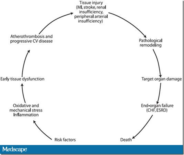
Another key principle endorsed by the HWG is that of the interrelationship between elevated BP and other CV risk factors. Even in patients with frank elevations in BP, risk stratification based on BP levels alone often underestimates CV risk. This is because above-optimal BP levels rarely occur in isolation, and patients seen in clinical practice frequently have multiple CVD markers or risk factors (eg, overweight, insulin resistance, dyslipidemia) that point to greater overall risk.[1,9-13] What is particularly significant, from the perspective of defining hypertension beyond BP thresholds, is that many of these disease processes are intimately interrelated and interact via common pathobiologic processes involving oxidative stress and endothelial dysfunction (Figure).[14] Moreover, the presence of risk factors and disease markers defines the earliest stage in this CVD continuum, well before overt CVD and target-organ damage can be measured in the clinic.[14] From this perspective, above-optimal BP (a risk factor) is not necessarily synonymous with hypertension (a disease representative of progressive CVD and tissue injury).[6]
image

Figure. Cardiovascular and renal pathophysiologic continuum. CV = cardiovascular; MI = myocardial infarction; CHF = congestive heart failure; ESRD = end-stage renal disease. Adapted from Dzau VJ, et al.[14]

Another consequence of the CV and renal pathophysiologic continuum is that the complex interplay of risk factors and disease markers frequently may manifest as a dramatically higher CV risk than would be expected, based on thresholds for each individual risk factor alone. This is highlighted by the particularly deleterious condition known as the cardiometabolic syndrome, in which individual risk factors combine to increase CV risk synergistically, rather than additively.[12,15] Ultimately, a more clinically meaningful assessment of CV risk can be obtained by global assessment of a patient's risk, rather than focusing solely on whether a patient has crossed a particular BP threshold.

Taken together, this evidence suggests that it may be more useful to view BP as 1, but not the only, biomarker for the disease hypertension, and to view above-optimal levels of BP in an individual patient as those that, when sustained, cause damage to the vasculature.[6] This forms the basis of the revised definition of hypertension, as shown in Table 1.

Table 1. Revised Definition of Hypertension From Hypertension Writing Group 2009

• Hypertension is a progressive CV syndrome arising from complex and interrelated etiologies
• Early markers of the syndrome are often present before BP elevation is sustained; therefore, hypertension cannot be classified solely by discreet BP thresholds
• Progression is strongly associated with functional and structural cardiac and vascular abnormalities that damage the heart, kidneys, brain, vasculature, and other organs, and lead to premature morbidity and death
• Reduction of elevated BP generally confers a reduction in the risk for CV events. Note that HWG separates elevated BP (one manifestation of the disease) from hypertension (the disease)

BP = blood pressure; CV = cardiovascular
From Giles T, et al.[6]

Because hypertension is defined by as "a progressive cardiovascular syndrome," it is clinically helpful to categorize, or stage, patients (Table 2), with each stage characterized by the cumulative presence or absence of markers of hypertensive CVD and evidence of target-organ damage. This provides a snapshot of the extent to which the disease has advanced at a particular time.[6]

Table 2. Revised Definition and Classification of Hypertension From Hypertension Writing Group 2009

Classification Normal Stage 1 Hypertension Stage 2 Hypertension Stage 3 Hypertension
Descriptive category Normal BP or rare BP elevations
AND
no identifiable CVD
Occasional or intermittent BP elevations
OR
early CVD
Sustained BP elevations
OR
progressive CVD
Marked and sustained BP elevations
OR
advanced CVD
Cardiovascular risk factors
(see Table 3)
None or few Several risk factors present Many risk factors present Many risk factors present
Early disease markers
(see Table 4)
None Usually present Overtly present Overtly present with progression
Target-organ disease
(see Table 5)
None None Early signs present Overtly present with or without CVD events

BP = blood pressure; CVD = cardiovascular disease
From Giles T, et al.[6]

In the broadest sense, individuals are classified as either normal or hypertensive based on their CV status (ie, the absence or presence of identifiable CVD), regardless of their BP pattern. The stages within the hypertension category are further refined, based on BP patterns or the extent of CVD (early, advanced, or progressive). Each hypertension stage is further characterized by the cumulative presence or absence of risk factors for CVD (Table 3);markers of hypertensive CVD, such as microalbuminuria or mild left ventricular hypertrophy (Table 4); and evidence of target-organ damage, such as frank albuminuria or moderate-to-severe left ventricular hypertrophy (Table 5).[6] The occurrence of a major cardiac event clearly places the progression of hypertensive CVD at a more advanced stage.[6]

Table 3. Cardiovascular Risk Factors

Increasing age
Elevated BPa
High heart rate
Overweight/obesity
    − Increased BMI
    − Central obesity
    − Increased abdominal circumference
    − Increased abdominal adiposity (waist-to-hip ratio)a
Dyslipidemia
    − Elevated LDL or non-HDLb cholesterol
    − Low HDL cholesterola
    − Elevated triglyceridesa
Elevated blood glucose, insulin resistance, or diabetes mellitusa
Chronic kidney disease
Smoking
Family history of premature CVD (age < 50 yr in men, < 60 yr in women)
Sedentary lifestyle
Psychosocial stressors
Elevated hs-CRP

BMI = body mass index; BP = blood pressure; CVD = cardiovascular disease; HDL = high-density lipoprotein; hs-CRP = high-sensitivity C-reactive protein; LDL = low-density lipoprotein
aComponents of the metabolic syndrome
bNon-HDL cholesterol = total cholesterol minus HDL cholesterol

Table 4. Early Markers of Hypertensive Cardiovascular Disease

System Physiologic Alteration
Blood pressure
Loss of nocturnal BP dipping
Exaggerated BP responses to exercise or mental stress
Salt sensitivity
Widened pulse pressure
Cardiac
Left ventricular hypertrophy (mild)
Increased atrial filling pressure
Decreased diastolic relaxation
Increased natriuretic peptide
Renal
Microalbuminuria (urinary albumin excretion of 30-300 mg/day)a
Reduced estimated GFR (60-90 mL/min)
Cerebrovascular
Stroke
Transient ischemic attack
Decreased cognitive function
Dementia
Loss of vision

BP = blood pressure; GFR = glomerular filtration rate
aAlso a marker of microcirculatory disease

Table 5. Hypertensive Target-Organ Damage and Overt Cardiovascular Disease

System Evidence of Target-organ Damage and Cardiovascular Disease
Cardiac
Left ventricular hypertrophy (moderate to severe)
Systolic or diastolic cardiac dysfunction
Symptomatic heart failure
Myocardial infarction
Angina pectoris
Ischemic heart disease or prior revascularization
Vascular
Peripheral arterial disease
Carotid arterial disease
Aortic aneurysm
Renal
Albuminuria (urinary albumin excretion > 300 mg/day)
Chronic kidney disease (estimated GFR < 60 mL/min) or ESRD
Cerebrovascular
Stroke
Transient ischemic attack
Decreased cognitive function
Dementia
Loss of vision

ESRD = end-stage renal disease; GFR = glomerular filtration rate

Clinical Characteristics and Practical Implications of the Proposed Hypertension Categories

A practical clinical interpretation of the revised hypertension categories is shown in Table 6.

Table 6. Clinical Characterization, BP Patterns, and Practical Implications of the Hypertension Algorithm

Hypertension Category Clinical Characterization BP Pattern Practical Implications


Normal
Optimal BP levels Resting BP levels usually < 120/80 mm Hg Includes some patients identified as having prehypertension (based on JNC 7 criteria)
No identifiable early markers of CVD Occasional BP elevations, even to ≥ 140/90 mm Hg, may occur


Stage 1
Early CVD markers present BP levels > 115/75 mm Hg Earliest identifiable stage of hypertensive disease
Frequently 1 or more CVD risk factors present BP may be frankly elevated, particularly with environmental stress Includes individuals with prehypertension (based on JNC 7 criteria) who also have CVD risk factors or early disease markers
No evidence of target-organ damage


Stage 2
Diffuse disease markers present
OR evidence (limited) of early target-organ damage
Sustained resting BP frequently ≥ 140/90 mm Hg, with much higher elevations induced by physiologic or psychologic stressors Equivalent to JNC 7 stage 1 hypertension
Indicates progressive disease
Risk factors, if not attenuated, continue to contribute to progressive target-organ disease


Stage 3
Overt CVD present Sustained resting BP levels ≥ 140/90 mm Hg usual (untreated or inadequately treated) Equivalent to JNC 7 stage 2 hypertension
Marked BP elevations to levels > 160/100 mm Hg not uncommon (untreated or inadequately treated) Includes all individuals with clinical evidence of overt target-organ damage or CVD, or who have sustained a CVD event, regardless of BP levels

BP = blood pressure; CVD = cardiovascular disease; JNC 7 = Seventh Report of the Joint National Committee on Prevention, Detection, Evaluation, and Treatment of High Blood Pressure

In the algorithm, individuals with optimal levels of BP and no identifiable early markers of CVD are categorized as normal. These individuals usually have resting BP levels of 120/80 mm Hg or lower, but BP may be elevated occasionally, even to levels of 140/90 mm Hg or higher.[6] Given the limits of clinic BP measurements, home BP determinations or 24-hour ambulatory BP recordings may be helpful in identifying patients with more than occasional BP elevations, who may be categorized more appropriately as having stage 1 hypertension.[16]Because the HWG algorithm does not recognize a prehypertension category, some individuals designated as having prehypertension according to the JNC 7 classification may be considered normal in the paradigm.

The earliest identifiable stage of hypertensive disease, stage 1 hypertension, is characterized by the presence of early CVD markers. Although BP levels are higher than 115/75 mm Hg and may be frankly elevated in patients at this stage, abnormal BP patterns -- including loss of nocturnal dipping, exaggerated responses to exercise or mental stress, and widened pulse pressure -- may provide clearer evidence of the presence of early hypertensive disease.[4] Although patients should have more than 1 CV risk factor to be included in this category, they should not have any evidence of target-organ damage.

In contrast to stage 1, stage 2 hypertension is characterized by diffuse disease markers and evidence of progressive disease as a consequence of persistent functional and structural changes in BP control mechanisms and in the heart and vasculature. Although patients at this stage frequently have sustained elevations in resting BP levels of 140/90 mm Hg or higher -- with much higher elevations induced by physiologic or psychologic stress -- it is important to recognize that any individual with numerous disease markers or limited evidence of early target-organ damage, such as left ventricular hypertrophy, fits into this category, regardless of BP levels. Methods of detecting or measuring some of the early target-organ damage characteristic of this stage of hypertension are currently limited to specialized or research settings, and further evaluation is needed to determine their potential utility and cost-effectiveness in clinical settings.[6] Nonetheless, aggressive management of CV risk factors that are identified in patients at this stage may help attenuate the progression of target-organ damage.

Finally, stage 3 hypertension is an advanced stage of the hypertensive continuum, characterized by the presence of overt CVD. Overt hypertensive target-organ disease is often pervasive, and CVD events may have already occurred. If inadequately treated or left untreated, individuals at this stage usually have sustained resting BP levels of 140/90 mm Hg or higher, although marked elevations to levels higher than 160/100 mm Hg are not uncommon.[6] Regardless of BP levels, however, all individuals with clinical evidence of overt target-organ damage or CVD, as well as those who have already sustained CVD events, are included in this category. Reaching this phase means that damage to target organs, as well as overt cardiorenal disease, has already occurred. As a consequence, CV risk factor modification and treatment of target-organ disease and all identified CVD should be vigorous and sustained.[6]

Strategies for and the Clinical Implications of Treating Patients Along a Continuum of Global Cardiovascular Risk

The paradigm shift in viewing elevated BP as a marker for hypertension and hypertension as a progressive CVD syndrome has important implications for treating patients in the clinical setting. The risk-based approach proposed by the HWG will lead to reclassifying patients who were previously designated prehypertensive (based on JNC 7 criteria) to either HWG normal or stage 1 hypertension.[4] In terms of treatment, lowering BP remains an important goal of antihypertensive therapy, yet ultimately the overarching objective is to prevent CV complications.[9] Treatment of other CV risk factors is therefore equally important. Moreover, CV risk factors, including elevated BP, are not only precipitators, but also continuous pathogenic components at every stage of progression of CVD.[9] Clinical strategies, therefore, need to focus on detecting and treating patients at risk at every stage along the continuum, from preventing target-organ damage and interrupting CVD progression in patients with early-stage hypertension, to making aggressive efforts to slow further disease progression and avoid CV events in patients with late-stage hypertension.

Evidence for the benefit of antihypertensive treatment in early-stage hypertension (HWG stage 1 or JNC 7 prehypertension category) has only recently become available. The Trial of Preventing Hypertension (TROPHY) study has shown that antihypertensive therapy may help prevent the development of elevated BP levels among individuals with BP lower than 140/90 mm Hg who are at high risk for frank hypertension (due to the presence of multiple CV risk factors).[17] In line with the HWG paradigm, patients in this study had high-normal SBP/DBP levels of 130-139/85-89 mm Hg at baseline, yet had a strikingly high rate of CV risk factors other than elevated BP.[17] Among the TROPHY patients, 96% had at least 1 additional CV risk factor, includingvarious measures of dyslipidemia, insulin resistance, and obesity, as well as elevatedhematocrit and heart rate; 81% had 2 or more additional risk factors; and33% had 4 or more additional risk factors. The most prevalent risk factor in thecohort as being overweight.[2,17] Patients were randomized to receive treatment with the angiotensin receptor blocker candesartan or placebo for 2 years, followed by an additional 2 years of placebo-only therapy; all patients were instructed to make changes in lifestyle to reduce BP throughout the trial.[17] After 2 years, hypertension haddeveloped in 154 patients in the placebo group and 53 patients in the angiotensin receptor blockergroup, representing a significant 63% relative risk reduction with pharmacotherapy (P < .001). After 4 years, hypertension had developed in 240 patients assigned to placeboand 208 patients assigned to active treatment (relative risk reduction, 15.6%;P < .007). Serious adverse events occurred in 3.5% of patients who received active treatment and in 5.9% of those who received placebo. As the authors noted, the absolute difference between active treatment and placebo at 2 years in TROPHY, 26.8%, is much higher than the 8% absolute difference observed in the Trials of Hypertension Prevention,[18] the only trial of lifestyle modification with a similar duration, suggesting that drug therapy plus lifestyle modification is more effective than lifestyle modification alone in early hypertension.[17]

The benefit of treatment with antihypertensive agents in patients classified as normotensive by conventional standards also is supported by the Perindopril Protection Against Recurrent Stroke Study (PROGRESS).[19] In this study, antihypertensive treatment in subjects without elevated BP (mean BP, 136/79 mm Hg) but with a history of target-organ damage -- in this case, a history of transient ischemic attacks or stroke -- was associated with a significant 27% reduction in the relative risk for stroke compared with placebo (P < .01), similar to the 32% reduction observed in patients designated as hypertensive.[19] Moreover, intensive BP reduction with combination therapy was associated with better outcomes than less-intensive BP reduction with single-agent therapy, regardless of hypertension status. Because individuals similar to those considered normotensive in PROGRESS would be classified as having stage 2 hypertension (based on the presence of target-organ damage) in the HWG paradigm, results of this study highlight the importance of considering comprehensive risk factor assessment, including the presence of target-organ damage, when defining and staging patients with hypertension.

Whether all individuals with early-stage hypertension, as defined by the HWG, should be treated with antihypertensive therapy requires further study. As the group emphasized in their 2005 report, characterizing hypertension as a complex CV disorder associated with, but not exclusively defined by, high BP is best viewed as a transitional strategy that is intended to generate further clinical research into improved strategies for detecting, treating, and possibly preventing the disease.[4]

Summary

The key points advanced by the HWG in their updated hypertension position paper are that BP serves as a biomarker for the disease hypertension and, as such, elevated BP is not synonymous with hypertension. Some individuals may exhibit elevated BP in the absence of hypertension, whereas other individuals with the same levels of BP might be classified into different stages of hypertension.[6] Therefore, for purposes of calculating total CV risk and staging patients as normal or hypertensive, BP should be evaluated in the context of other CV risk factors and disease markers. Ultimately, it is hoped that the risk-based approach to defining and staging hypertension, as proposed by the HWG, will lead to earlier identification of individuals with hypertensive CVD. Preliminary data, such as that described by the TROPHY Investigators, suggest that lowering BP with pharmacologic therapy can prevent or delay the progression of hypertensive CVD even at early stages (ie, HWG stage 1 hypertension/JNC 7 prehypertension). Additional research is necessary to confirm these findings and identify cost-effective methods to detect and measure early CVD markers in clinical practice.

This activity is supported by an independent educational grant from Pfizer.

This article is a CME certified activity. To earn credit for this activity visit:
http://cme.staging.medscape.com/viewarticle/708548

References
  1. Weir MR. Risk-based classification of hypertension and the role of combination therapy. J Clin Hypertens (Greenwich). 2008;10:4-12.
  2. Giles TD. Assessment of global risk: a foundation for a new, better definition of hypertension. J Clin Hypertens (Greenwich). 2006;8:5-14.
  3. Khosia N, Black HR. Expanding the definition of hypertension to incorporate global cardiovascular risk. Curr Hypertens Rep. 2006;8:384-390.
  4. Giles TD, Berk BC, Black HR, et al. Expanding the definition and classification of hypertension. J Clin Hypertens (Greenwich). 2005;7:505-512.
  5. Chobanian AV, Bakris GL, Black HR, et al; National Heart, Lung, and Blood Institute Joint National Committee on Prevention, Detection, Evaluation, and Treatment of High Blood Pressure; National High Blood Pressure Education Program Coordinating Committee. The Seventh Report of the Joint National Committee on Prevention, Detection, Evaluation, and Treatment of High Blood Pressure: the JNC 7 report. JAMA. 2003;289:2560-2572.
  6. Giles TD, on behalf of the Hypertension Writing Group. Definition and classification of hypertension: an update. J Clin Hypertens (Greenwich). In press.
  7. Sierra C, de la Sierra A. Early detection and management of the high-risk patient with elevated blood pressure. Vasc Health Risk Manag. 2008:4;289-296.
  8. Franklin SS, Khan SA, Wong ND, et al. Is pulse pressure useful in predicting risk for coronary heart disease? The Framingham Heart Study. Circulation. 1999;100:354-360.
  9. Basile J. Management of global risk across the continuum of hypertensive heart disease. J Clin Hypertens (Greenwich). 2006;8:21-30.
  10. Khot UN, Khot MB, Bajzer CT. Prevalence of conventional risk factors in patients with coronary heart disease. JAMA. 2003;290:898-904.
  11. Neaton JD, Wentworth D; the Multiple Risk Factor Intervention Trial Research Group. Serum cholesterol, blood pressure, cigarette smoking, and death from coronary heart disease: overall findings and differences by age for 316,099 white men. Arch Intern Med. 1992;152:56-64.
  12. Meigs JB, D'Agostino RB Sr, Wilson PW, et al. Risk variable clustering in the insulin resistance syndrome: the Framingham Offspring Study. Diabetes. 1997;46:1594-1600.
  13. Gregg EW, Cheng YJ, Cadwell BL, et al. Secular trends in cardiovascular disease risk factors according to body mass index in US adults. JAMA. 2005;293:1868-1874.
  14. Dzau VJ, Antman EM, Black HR, et al. The cardiovascular disease continuum validated: clinical evidence of improved patient outcomes: Part I: pathophysiology and clinical trial evidence (risk factors through stable coronary artery disease). Circulation. 2006;114:2850-2870.
  15. Hoerger TJ, Ahmann AJ. The impact of diabetes and associated cardiometabolic risk factors on members: strategies for optimizing outcomes. J Manag Care Pharm. 2008;14:S2-S14.
  16. Pickering TG, White WB; American Society of Hypertension Writing Group. ASH position paper: home and ambulatory blood pressure monitoring. When and how to use self (home) and ambulatory blood pressure monitoring. J Clin Hypertens (Greenwich). 2008;10:850-855.
  17. Julius S, Nesbitt SD, Egan BM, et al; Trial of Preventing Hypertension (TROPHY) Study Investigators. Feasibility of treating prehypertension with an angiotensin-receptor blocker. N Engl J Med. 2006;354:1685-1697.
  18. The Trials of Hypertension Prevention Collaborative Research Group. Effects of weight loss and sodium reduction intervention on blood pressure and hypertension incidence in overweight people with high-normal blood pressure: the Trials of Hypertension Prevention, phase II. Arch Intern Med. 1997;157:657-667.
  19. PROGRESS Collaborative Group. Randomised trial of a perindopril-based blood-pressure-lowering regimen among 6,105 individuals with previous stroke or transient ischaemic attack. Lancet. 2001;358:1033-1041.
Read More .......